
Can a hip feel “tight” or painful because a ligament is loose rather than shortened? That question matters because the hip's stabilizing tissues don't all restrain the same movement. A symptom's location can be useful, but the position that brings it on often tells a clinician more about which structures may be involved.
The ligaments in the hip work with the joint capsule, muscles, labrum, and the shape of the bones to steady the joint while it moves. Understanding which tissues resist extension, rotation, or abduction can help make sense of symptoms, but it can't diagnose an injury on its own.
Table of Contents
- Hip Ligaments Reference Chart
- The Iliofemoral Ligament, the Hip's Strongest Restraint
- Which Ligament Restrains Which Hip Movement
- Ligamentum Teres and Zona Orbicularis as Stabilizers
- How Hip Ligaments Load During Walking
- Ligament Injury and Laxity Signs to Recognize
- What Hip Ligament Health Means for Daily Mobility
- Confirm the Cause Before Choosing Treatment
Hip Ligaments Reference Chart
The hip has three principal capsular ligaments, plus two other structures that matter clinically: the circumferential zona orbicularis and the ligamentum teres. The table is a quick reference to where each sits and what it helps restrain.
| Structure | Attachments | Main restriction | Clinical note |
|---|---|---|---|
| Iliofemoral ligament, or Y ligament of Bigelow | Anterior inferior iliac spine to the intertrochanteric line of the femur | Limits extension and external rotation | Dominant anterior restraint; resists anterior translation of the femoral head |
| Pubofemoral ligament | Pubic region to anterior femur | Limits excessive abduction and extension | Medial anterior capsule; restrains abduction in extension |
| Ischiofemoral ligament | Posterior acetabular region toward the femur | Limits internal rotation and adduction in flexion | Posterior capsular restraint; contributes to stability in FADIR-type positions |
| Zona orbicularis | Circular fibers around the femoral neck, without a broad bony attachment | Resists distraction in neutral and helps center the femoral head | Helps reduce translation, edge loading, and subluxation risk |
| Ligamentum teres | Fovea of the femoral head to the acetabular notch and transverse ligament | Contributes to rotational stability | Inside the joint, not part of the capsule; easy to overlook |
How to read the chart
The first three rows describe capsular ligaments. They aren't isolated cords, but spiral-shaped reinforcements woven into the capsule. Their orientation helps them tighten in certain hip positions, so a ligament's role depends partly on how the hip is moving.
The other two rows describe structures with different locations and jobs. The zona orbicularis is a circular band of capsular fibers around the femoral neck. The ligamentum teres sits inside the joint, connecting the femoral head to the acetabular notch and transverse ligament.
Match the structure to the symptom pattern
For symptoms at the front of the hip, the iliofemoral and pubofemoral ligaments are relevant because they help restrain movement at the anterior capsule. Posterior symptoms or discomfort in a flexed, inward-rotated position may involve the ischiofemoral region, though muscles and other joint structures can produce similar sensations.
A feeling of slipping or giving way raises a broader stability question. The zona orbicularis, ligamentum teres, capsule, and surrounding muscles can all contribute to control, so the name of one ligament rarely explains the full picture. The following sections examine how these structures respond to specific positions and why that matters during movement.
The Iliofemoral Ligament, the Hip's Strongest Restraint
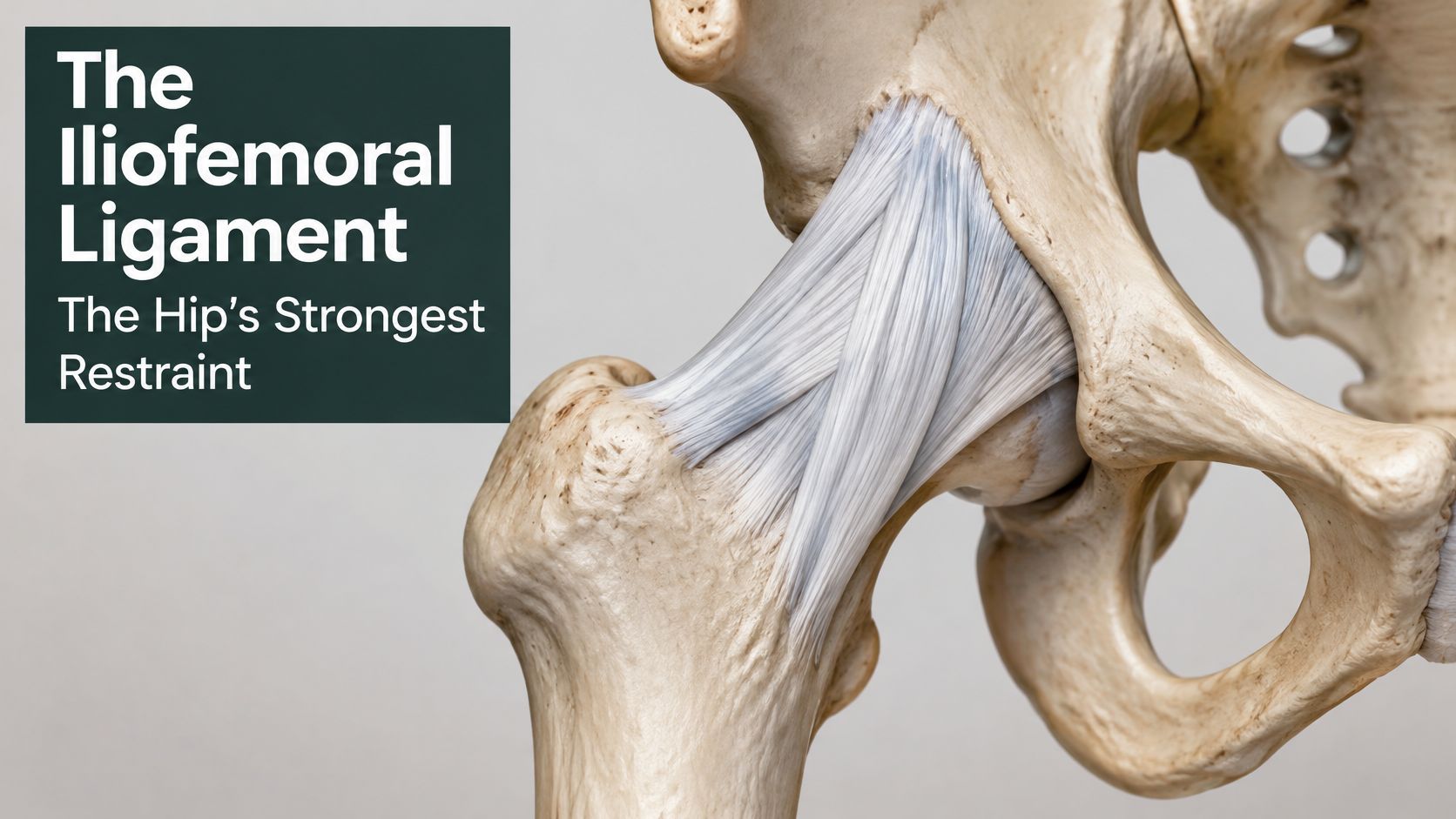
The iliofemoral ligament is described anatomically as the strongest ligament in the human body. Also called the Y ligament of Bigelow, it runs from the anterior inferior iliac spine to the intertrochanteric line of the femur. Its two arms, commonly described as medial and lateral, reinforce the front of the hip capsule and help limit extension and external rotation.
That inverted-Y shape explains a lot about how it behaves. The apex anchors at the anterior inferior iliac spine, and the two arms spread downward to attach along the intertrochanteric line, so the ligament drapes across the front of the joint rather than crossing it as a single narrow cord. Because the fibers fan out, part of the structure can stay under some tension across a wider arc of positions than a thin band would manage.
Why extension puts it under tension
The ligament's spiral orientation helps it tighten as the hip extends. That tension resists excessive forward movement of the femoral head and supports the joint in an upright position. In practical terms, the ligament helps a person stand with the hip extended without requiring the surrounding muscles to work constantly to hold the joint steady.
A history that fits this restraint often involves the trunk rather than the leg. Think of a patient who feels apprehension at the front of the hip when leaning backward, whether tipping the head back to rinse hair in a shower or arching to look up while standing. Leaning the trunk back is relative hip extension, and it shifts the femoral head toward the front of the socket, raising demand on the anterior capsule. Adding outward rotation to that lean tightens the arms of the Y further still. A symptom that tracks those positions is worth describing precisely during an assessment, even though the position alone cannot confirm which structure is irritated.
The same front-of-joint restraint helps explain why hip dislocations are predominantly posterior. Approximately 90% of hip dislocations are reported to be posterior, and the NCBI Bookshelf overview of hip anatomy describes the anterior reinforcement, especially from the iliofemoral ligament, that makes forward displacement the harder direction. For an individual, that anatomy supplies context for an examination rather than a verdict.
What a symptom description can tell you
“My hip hurts when I walk” does not identify a ligament. The details that narrow the picture are positional: whether pain appears as the leg passes behind the body, whether turning the leg outward reproduces it, and whether the joint feels unreliable rather than merely sore. Each answer steers a clinician toward different structures to examine, because muscles, the labrum, and joint shape can all imitate an anterior-capsule complaint.
Practical rule: Anterior pain brought on by extension and outward rotation is a reason to assess hip stability, not proof of a torn iliofemoral ligament.
When reading a symptom description, remember this: the iliofemoral ligament is the dominant front-of-hip restraint, and the position that provokes symptoms tells you more than the label “hip ligament pain.”
Which Ligament Restrains Which Hip Movement
Each capsular ligament tightens at a different joint angle, the way ropes around a ship's mast take load only when it leans in their direction. Extension tensions the front of the capsule, flexion with inward rotation loads the back, and a pull straight along the femoral neck is checked mainly by its ring-shaped fibers. This review of hip capsular anatomy and stability describes these position-dependent roles and explains why surgeons aim to preserve the capsule during procedures.
| Movement or position | Primary restraint | Supporting restraint |
|---|---|---|
| Extension | Iliofemoral ligament | Pubofemoral ligament |
| Extension with external rotation | Iliofemoral ligament | Pubofemoral ligament; zona orbicularis |
| Abduction in extension | Pubofemoral ligament | Iliofemoral ligament |
| Internal rotation in flexion | Ischiofemoral ligament | Zona orbicularis |
| FADIR, or flexion, adduction, and internal rotation | Ischiofemoral ligament | Iliofemoral ligament |
| Neutral distraction of the joint | Zona orbicularis | Capsular ligaments |
Anterior restraints in extension
A dancer holding an extended leg turned outward is loading the capsule in exactly the combination the front of the joint resists: extension with external rotation, made more demanding still if the leg is also drawn toward the midline. The same leg held in a flexed, seated posture places far less strain on those fibers.
The pubofemoral ligament takes over as the leg moves away from the body in extension. A martial artist driving into a high side kick combines abduction with a straightening hip, asking the lower front of the capsule to restrain a motion that would otherwise carry the femoral head outward.
Posterior restraint in flexion and rotation
The ischiofemoral ligament contributes to posterior stability during internal rotation and the combined position known as FADIR, meaning flexion, adduction, and internal rotation. Pulling a leg across the body while seated or moving into a deep yoga twist can approach that position. Pain during FADIR deserves careful interpretation because it can reflect posterior capsular loading, but it may also relate to other joint structures.
The zona orbicularis works on a different principle. Rather than spanning one direction of rotation, it forms a collar around the femoral neck that resists the joint being pulled apart in neutral and keeps the femoral head seated in the socket through extreme flexion and extension. Its job is centering the head, so it shares load in nearly every row of the table.
Why capsule preservation matters
A procedure that disrupts the capsule can weaken more than one restraint at once. Damage to the zona orbicularis or the iliofemoral ligament may compromise both rotational and translational stability, which is why clinicians weigh capsular preservation and the avoidance of excessive laxity when considering hip arthroscopy or another intervention.
A movement test narrows the list of suspects. Pain location, strength, joint range, and whether symptoms resemble a labral or muscle-tendon problem complete the picture before anyone assigns blame to a single ligament.
Ligamentum Teres and Zona Orbicularis as Stabilizers
A hip can feel unstable in one position yet controlled in another because its internal ligaments and capsule share the work of guiding the joint. The ligamentum teres sits inside the joint, connecting the femoral head to the hip socket. A 2025 analysis of ligamentum teres and capsular function describes its contribution to rotational stability in the frontal and transverse planes, with its role changing according to hip position.
The zona orbicularis has a different stabilizing role: its circular fibers help resist the femoral head being pulled away from the socket. These structures can contribute to stability, but anatomy alone cannot identify the source of pain. Symptoms near the joint may also involve the labrum or capsule, so assessment needs to consider which movement brings them on.
A computational study of total hip arthroplasty found that, in its model, the capsule supplied an average of 31.0% of resistance to internal-external rotation, while passive local muscles supplied 69.0%. The model also estimated that the iliofemoral ligament contributed up to 80.8% of resistance to external rotation in hyperextension. These figures describe a replaced hip model, so they should not be applied directly to a natural hip.
Muscles and ligaments work together to control movement. Unstable hips often need better control more than extra end-range mobility. Pregnancy, prior surgery, or generalized hypermobility may affect how a position feels, but those factors alone do not establish which tissue is causing pain.
How Hip Ligaments Load During Walking
A biomechanical analysis of hip ligament loading during gait reported mean peak strains of about 9.5% for the iliofemoral ligament at 51.5% of the gait cycle, 5.7% for the pubofemoral ligament at 57.4%, and 4.7% for the ischiofemoral ligament at 35.5%; the same work reported cadaveric capsule-ligament tensile strength around 1.65 MPa and elastic modulus around 14.23 MPa. Those laboratory figures describe tissue mechanics. They do not set an injury threshold for a living hip.
What the gait-cycle points mean
The gait cycle starts when one foot contacts the ground and ends when that same foot contacts the ground again. The iliofemoral peak lands in late stance, when the leg is behind the body and the hip is extending just before toe-off. A useful analogy is a seat belt that tightens only when the car reaches a certain angle: the ligament matters most when hip position pulls it taut.
| Ligament | Peak timing during stance | Why that timing matters | Position that lowers strain |
|---|---|---|---|
| Iliofemoral | Late stance, as the hip extends | Anterior restraint is loaded when the leg trails the body | Hip flexion; internal rotation plus adduction |
| Pubofemoral | Later in stance, after the iliofemoral peak | Anteroinferior restraint may share load near toe-off | No lowering position reported; use extension-provoked symptoms as a clue |
| Ischiofemoral | Earlier in stance | Posterior restraint is checked before the trailing-leg phase | No lowering position reported; FADIR-type symptoms guide posterior checks |
Apply the findings carefully
If anterior capsular involvement is suspected, a clinician may test pain, apprehension, or control during late-stance extension and during tasks that combine extension with outward rotation. FADIR-related symptoms can prompt evaluation of posterior capsuloligamentous loading while also raising the possibility of an intra-articular source. The provoking position narrows the search; it cannot name the injured tissue by itself.
The practical cue is timing. Pain near the end of a step points attention toward structures loaded in extension, while symptoms earlier in stance suggest a different restraint pattern. Match the complaint to the phase that provokes it, then confirm the tissue with examination rather than assuming a generic hip ligament problem.
Ligament Injury and Laxity Signs to Recognize
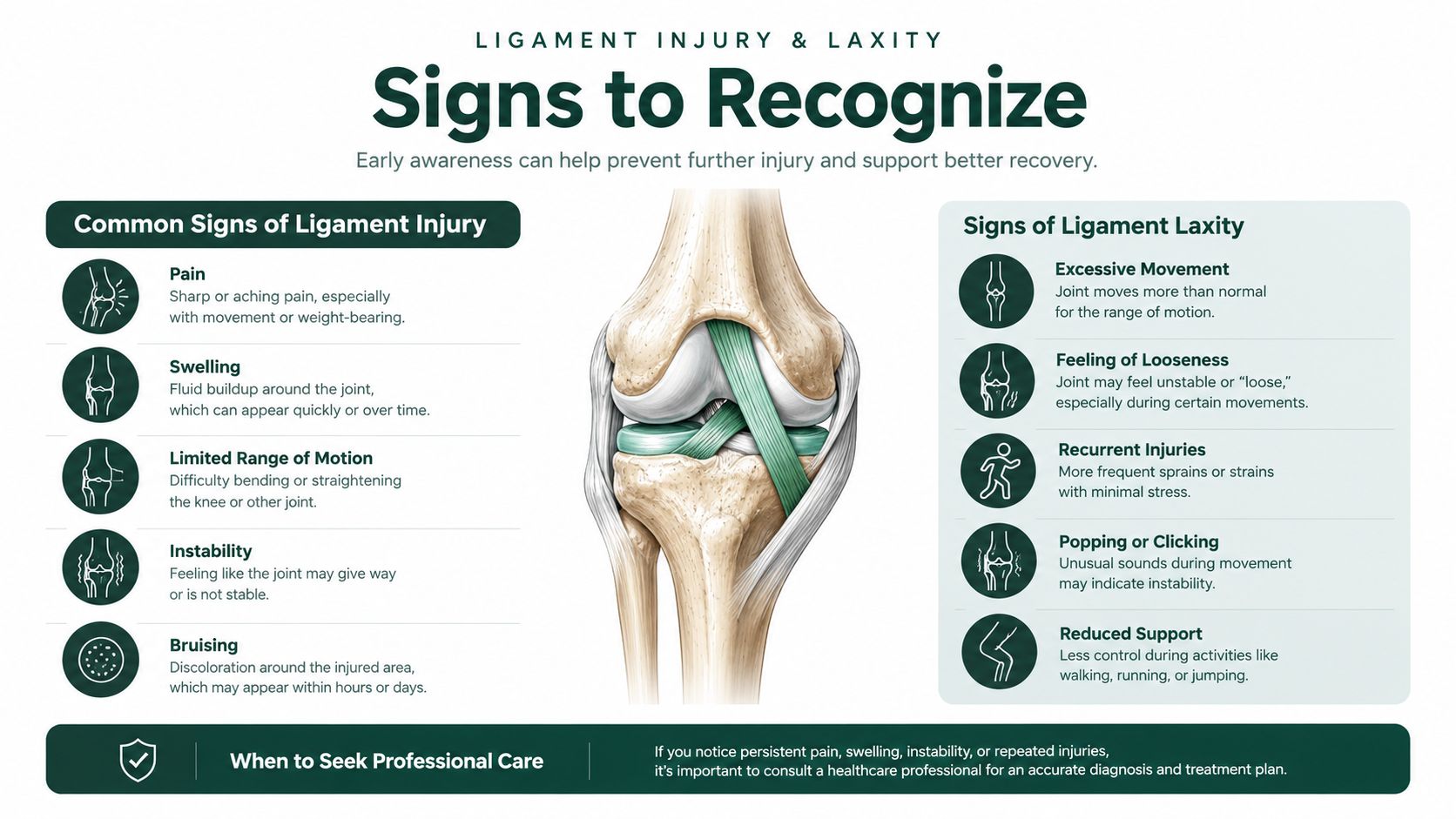
Hip and groin problems are common in sport, but the published injury figures are not counts of ligament tears. A systematic review and meta-analysis of hip and groin injuries in sport covering 71 studies, 5,914 injuries, and 9,441,381 hours of athletic exposure found an overall incidence of 0.71 injuries per 1,000 exposure hours, with hip and groin problems representing 11% of reported sports injuries. Adductor-region injuries accounted for about 6% of all injuries, and the estimated burden was 11.5 days lost per 1,000 exposure hours. Those figures group muscle, tendon, and joint-structure injuries together, so they describe the functional burden of the region rather than how often a ligament alone is injured.
Clues that may point toward instability
Instability complaints follow a pattern worth learning: the position that provokes the symptom points to the restraint under suspicion. A review of clinical clues to hip microinstability found anterior hip pain in 78% of cases, a subjective sense of instability while walking in 81%, and apprehension or pain with hip extension combined with external rotation in 65%. That third clue is the position-specific one. Extension plus outward rotation loads the anterior capsule, where the iliofemoral ligament does most of its restraining work, so apprehension in that position raises suspicion of anterior capsular involvement rather than a generic hip ligament problem.
These numbers are clues, not diagnostic rules. The review included only nine studies and 189 patients, and every included study was level-IV evidence.
Ligaments behave like straps that tighten only near the end of a movement. A strap that feels loose in the middle of the range may still hold firmly where it counts, which is why laxity alone isn't a diagnosis: a person can be naturally flexible and still have good function. What justifies a focused stability assessment is the pattern. Repeated giving way, pain or apprehension when the hip reaches extension and outward rotation, or a recurring sense that the joint is slipping during that specific task all carry more weight than looseness on its own.
Hip pain can also arise from the labrum, hip-flexor tendons, pelvic-floor dysfunction, or the lumbar spine. If labral symptoms are part of the concern, this guide to understanding recovery after a torn labrum offers additional context. A clinician should consider strength, range of motion, movement mechanics, and the whole kinetic chain rather than treating every front-of-hip symptom as a ligament problem.
What Hip Ligament Health Means for Daily Mobility
Hip ligaments provide passive stability. Each one acts as a restraint for a particular joint position, so in daily life the informative detail is which movement provokes symptoms. Walking, sitting, squatting, and turning each load a different part of the capsule, and muscles supply active control on top of what the passive tissues provide.
Pregnancy and postpartum movement
Pregnancy can bring postural changes that alter how a person loads the hip and pelvis. For a new parent carrying a baby on one side, repeated weight shifts challenge the muscles and soft tissues that help control the joint. Noticing which activities provoke symptoms, and whether control fades as fatigue builds, gives a clinician more to work with than a general report of hip pain.
Osteopathic care can address tension, discomfort, mobility issues, and musculoskeletal strain that often accompany pregnancy and parenting. A whole-body approach to osteopathic manipulative treatment can help readers understand how a clinician may consider movement beyond one painful spot.
Sport and flexibility
High-load or fast movement asks the hip to manage force while changing direction. A hiker descending a steep trail repeatedly reaches hip extension as the leg trails behind, the position the iliofemoral ligament restrains. A tennis player planting and pivoting loads several restraints at once. If pain or apprehension shows up in the same position again and again, that pattern points toward a restraint problem worth examining.
Someone with extreme natural range and no symptoms needs no intervention. Another person with the same range who feels pain every time the leg trails far behind presents a different clinical question, because the symptom is tied to a specific restraint rather than to flexibility itself.
Support useful movement
Practical support for hip stability means training the movements the hip performs in daily life:
- Gluteus medius and rotator control: Build coordination in the muscles that keep the pelvis level and the femoral head centered during single-leg tasks like stepping off a curb.
- Progressive loading: Move from level walking to hills and stairs, then to deeper squats, before attempting demanding end-range positions.
- End-range tolerance: Develop control in the specific positions a task requires, such as the deep flexion of tying a shoe or the extension of a long stride.
- Symptom-free patterns: Check that ordinary movement stays comfortable and controlled across the day.
When a surgeon operates on the labrum or capsule, it's reasonable to ask whether the capsule was preserved, closed, or plicated. That question matters because capsular tissue contributes to joint stability, and the appropriate surgical choices depend on the person's anatomy and clinical needs.
Confirm the Cause Before Choosing Treatment
Ligament-related microinstability is a stability problem, not just a pain or tightness diagnosis. A clinician considers symptoms alongside examination, hip morphology, capsule condition, and muscular control. Imaging can be inconclusive, and one MRI finding by itself doesn't establish functional instability.
Use these clues to describe your experience, not to diagnose yourself:
- The hip gives way during walking.
- Extension is painful, particularly when the leg turns outward.
- You feel apprehension in certain positions or a repeated sensation that the joint is slipping or subluxing.
- Clicking or catching occurs with deep flexion, which may point toward a labral or other joint concern.
- Pain appears when you resist hip flexion, which may suggest a hip-flexor tendon problem.
- Pelvic-floor symptoms or pain referring from the lower back may indicate another source.
A focused appointment should assess the whole kinetic chain and distinguish ligament concerns from labral, tendon, pelvic-floor, and lumbar causes. Bring a short log noting which positions or activities provoke symptoms, when they occur, and any history of dislocation, pregnancy, or surgery. A review of clinical clues to hip microinstability emphasizes that the available evidence is limited, so these signs should guide evaluation rather than be treated as proof.
Practical next steps
- Keep a symptom log until your first appointment. Note the movement, timing, and whether the hip feels painful, apprehensive, or unstable.
- Book a clinical assessment that includes movement-specific testing and considers other possible pain sources.
- Discuss rehabilitation focused on movement control and progressive loading. A clinician may also consider osteopathic manipulative care or regenerative injection therapy after assessment. Regenerative injection therapy may be evaluated for joint pain in the knees, shoulders, or hips, ligament and tendon injuries, chronic back or neck pain, arthritis, and joint instability. Prolotherapy involves injecting a solution, commonly containing a dextrose-based proliferant, into targeted musculoskeletal tissues. Learn more about regenerative injection therapy.
Sudden inability to bear weight, a visibly out-of-place hip, or numbness calls for urgent care, not a routine appointment.
Restore Osteopathic Medicine offers osteopathic care for mobility and musculoskeletal concerns, along with evaluation of regenerative injection therapy for selected joint and ligament conditions. If hip pain or instability is affecting your daily movement, visit Restore Osteopathic Medicine to learn about care options and request an evaluation.