
What if a ligament looks thick on a scan taken while you're lying flat, but the actual trouble only shows up when you stand and lean back? That question sits at the heart of buckling of ligamentum flavum. It's a finding that puzzles many people, since a report may describe a thickened ligament, yet the symptoms rise and fall with posture. Those two facts fit together without conflict. A ligament can fold inward in one position and lie flatter in another, so a single image may not capture the full picture.
Table of Contents
- Buckling of Ligamentum Flavum at a Glance
- How Disc Loss and Extension Make the Ligament Fold
- Why Lying-Down Scans Miss Buckling and Why It Is Not Just Thickness
- Symptoms That Point Toward Buckling
- Treatment Options Matched to the Mechanical Problem
- Your Next Step With Suspected Buckling
Buckling of Ligamentum Flavum at a Glance
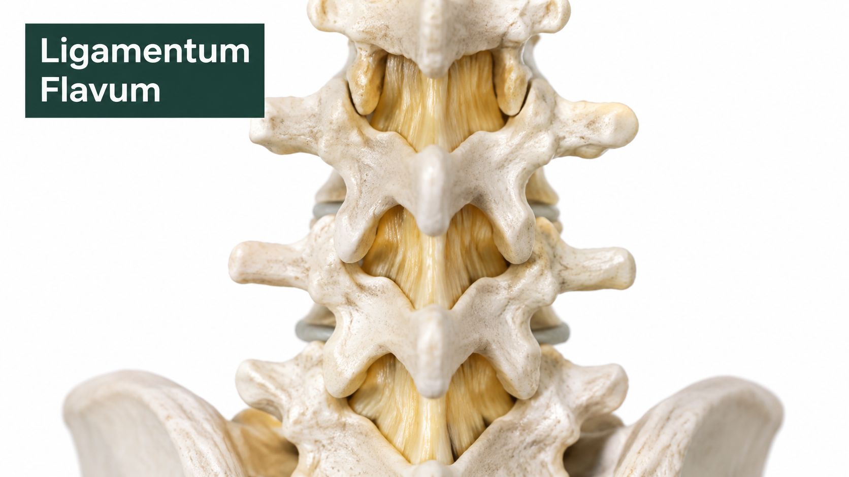
Buckling of ligamentum flavum is the inward folding of the yellow, elastic ligament that connects neighboring laminae at the back of the spine into the spinal canal, usually when the back extends.
Think of the ligamentum flavum as a tough elastic strap. It lines the back wall of the canal, helps protect the nerves inside, and resists excessive motion between vertebrae. When it's healthy and taut, it stays out of the way. When it has slack, or when it's already thickened, it can bulge inward and leave the nerves less room.
Key figures for ligamentum flavum thickness and buckling
| Measure | Value | What It Means |
|---|---|---|
| Ligament thickness in controls at L4-L5 | 2.25 ± 0.51 mm | Reference range for participants without central stenosis |
| Ligament thickness in central stenosis at L4-L5 | 4.02 ± 0.74 mm | About 1.77 mm thicker, roughly 79% greater than controls |
| Diagnostic cutoff for stenosis | 3.10 mm, 95.0% sensitivity, 94.2% specificity, AUC 0.97 | Strong separation of stenosis from controls, but a thickness value alone does not give the mechanism |
| Apparent thickness in extension | About 4 mm rising to over 9 mm in some patients | Shows that buckling changes the ligament's apparent size with posture |
Those numbers come from an observational MRI study in the Medicine journal article on ligament thickness at L4-L5, which compared 101 participants with central lumbar spinal canal stenosis with 103 controls and found an average thickness of 4.02 ± 0.74 mm in the stenosis group versus 2.25 ± 0.51 mm in controls.
Read the first two rows together. They don't describe a tumor-like growth. They describe a ligament with less reserve space around it, so even a small inward fold matters more. The cutoff row shows thickness separates groups well, but it doesn't tell you whether you're seeing fixed enlargement, a fold, or both. The last row is the clue that posture changes the picture. The sections that follow explain why the ligament folds, why lying-down scans can miss it, what symptoms fit, and what treatments can and can't do.
How Disc Loss and Extension Make the Ligament Fold
As the disc loses height, the space between the vertebrae changes, leaving the ligamentum flavum with more slack. When the lower back extends, the bony edges behind the spinal canal move closer together, and that slack can fold inward.
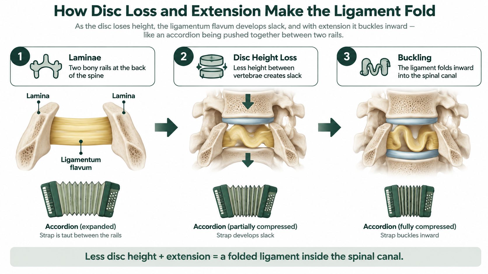
The sequence is straightforward:
- Disc height drops. As a disc loses height with degeneration, the gap between the laminae, the bony plates at the back of the vertebrae, can shrink. The ligament spans a shorter distance and has more slack.
- The back extends. Standing upright or arching backward can bring the laminae closer still.
- The ligament folds inward. The slack tissue can buckle toward the spinal canal rather than lying taut along its back wall.
- The canal narrows. The fold takes up room used by the nerve sac.
- Nearby structures have less space. The fold can displace epidural fat and reduce room for nerve roots.
A geometric problem first
Disc-height loss and movement can change the ligament's shape and position. The StatPearls chapter on lumbar spinal stenosis describes how disc-height loss can leave the ligament relatively redundant. This helps explain why a thick-looking ligament on one scan may reflect a posture-dependent fold rather than a fixed diagnosis.
Disc changes can occur alongside other sources of narrowing. A guide to an S1 bulging disc explains the disc component. Facet hypertrophy, osteophytes, and spondylolisthesis may also be present. Each can reduce available space, so several findings together may contribute to canal narrowing.
The tissue itself may also change with degeneration. One proposed explanation is that changes in elastic fibers and stiffer material affect how readily the ligament recoils as its attachment points shift. These tissue changes and the ligament's position can contribute to the same fold, while other nearby structures may add to the narrowing.
Why Lying-Down Scans Miss Buckling and Why It Is Not Just Thickness
Can a ligament look thick on an MRI because it has permanently enlarged, or because its shape changes with your posture? The answer matters: buckling is a position-dependent fold, and a scan taken while lying down may not show how the ligament behaves when the spine extends.
A supine MRI captures the spine at rest, often with the knees slightly supported. In that position, the ligament may lie flatter. Standing, walking, or arching backward can change the space around it, so a resting scan may understate the narrowing that occurs during those activities.
Detection of buckling by imaging position
| Imaging Approach | Patient Position | Buckling Detected | Neural Compression Seen |
|---|---|---|---|
| Conventional supine MRI | Lying flat | 9% of participants | Less conspicuous on routine imaging |
| Dynamic upright or extension MRI | Standing or extended | 80% of participants | 87% of patients |
In the dynamic MRI study published in the Egyptian Journal of Radiology and Nuclear Medicine, standard MRI identified ligamentum flavum buckling in 9% of participants, compared with 80% on upright or extension-based imaging. The study also found that buckling tended to occur alongside disc protrusion and foraminal narrowing, suggesting the fold can be part of a broader pattern of reduced space.
A thick-looking ligament does not always mean the tissue itself has enlarged. True hypertrophy is an increase in tissue; buckling is existing tissue folding inward as the spine's position changes. Both can occur together, and one lying-down image may not distinguish them clearly. Disc-space narrowing and segmental instability can contribute to the change in shape.
Buckling is a mechanical fold, rather than a sign of muscle tightness or inflammation. Nearby muscles may ache or guard, and nerves may become irritated, but those responses do not explain why the ligament folds.
An MRI finding alone does not determine treatment. The imaging needs to match the person's symptoms, examination, and the spinal level involved. A striking image at a level that does not fit the clinical picture may not explain the problem.
The question to ask: Can a neutral, lying-down MRI explain symptoms that happen mainly while standing, walking, or extending the spine?
When symptoms change with posture, that question may help a clinician decide whether additional imaging would be useful. Dynamic or upright imaging is not available everywhere and is not automatically needed. It may be considered when symptoms are strongly posture-dependent but the supine scan looks mild, or when imaging appears severe but does not fit the examination.
Positioning affects what the images show. How far the spine extends, how long that position is held, and how measurements are made can all influence the result. Even when dynamic imaging shows narrowing, the finding needs to match the symptoms and examination. It explains a possible mechanism, but does not by itself determine whether an injection, surgery, or neither is appropriate.
A thick ligament on one scan may reflect fixed enlargement, a posture-dependent fold, or a mixture of both. A normal-looking supine scan does not rule out buckling, while a visible fold on extension does not automatically call for a procedure. The key is whether the imaging, spinal level, and clinical findings fit together.
Symptoms That Point Toward Buckling
The classic pattern isn't constant pain. It's leg pain, heaviness, tiredness, or numbness that builds with standing and walking, then eases when you sit down or lean forward. Many people notice they can walk farther leaning on a shopping cart than walking upright. That posture-linked pattern, often called neurogenic claudication, fits dynamic canal narrowing well because sitting and flexing forward tend to open the canal and stretch the ligament flatter.
Symptom patterns and what they suggest
| Symptom Pattern | What It Points Toward | Next Question |
|---|---|---|
| Leg pain, heaviness or numbness that builds with standing and walking, and eases when sitting or leaning forward | Dynamic canal narrowing, the pattern seen in neurogenic claudication | Does a dynamic study show narrowing during extension at the matching level? |
| Pain or tingling triggered by arching backward and relieved by bending forward | Extension-sensitive encroachment on nerve space | Do the examination findings match the same side and level? |
| Constant pain at rest with night pain, fever or weight loss | Not typical for simple buckling | Are there other causes that need prompt evaluation? |
| Progressive leg weakness, numbness in the groin or saddle area, or new bladder or bowel changes | Possible severe compression | Does the person need emergency evaluation now? |
Read the rows in order. The first two describe symptoms that track with position, which is exactly what you'd expect when a fold appears in extension and flattens in flexion. Details help here. Note whether symptoms hit one leg or both, how far you can walk before they start, and how quickly sitting brings relief.
The third row is a caution against anchoring on the ligament. Constant rest pain, night pain, fever, or unexplained weight loss don't fit simple buckling and need prompt evaluation for other causes. Don't let an MRI phrase explain away a pattern it doesn't match.
The fourth row is urgent. Progressive leg weakness, numbness in the groin or saddle area, or new bladder or bowel changes can signal severe compression and call for emergency medical evaluation. Don't wait for a routine appointment if those appear.
Symptoms suggest, they don't confirm. Buckling is a reasonable explanation only when the level, side, and neurological examination match the pattern you describe.
Other conditions can mimic parts of this picture, including hip and pelvic problems. A resource on how to treat sacroiliac joint pain illustrates why a careful examination sorts out overlapping pain sources instead of relying on symptoms alone. A simple log recording activity, posture, and timing gives your clinician the fluctuating pattern that's often the most informative clue of all.
Treatment Options Matched to the Mechanical Problem
Start with a caution that protects you from overpromises. The evidence doesn't establish that manual therapy or regenerative treatments reverse buckling itself. Anyone claiming to unfold or shrink the ligament with a hands-on technique or injection is going beyond what's shown. That doesn't mean conservative care is useless. It means its realistic target is the mechanical environment around the ligament, not the fold in isolation.
It's also worth knowing the ligament isn't expendable packing material. It's part of the posterior tension band and contributes to spinal stability and elastic recoil. The review on ligamentum flavum mechanics and treatment considerations notes that a 2026 ex-vivo lamb-spine study found that total ligamentum flavum removal increased motion and reduced stiffness under multiple loading conditions. That's an animal laboratory finding, not a human outcome, but it supports treating the ligament as a stabilizing structure when removal is discussed.
Treatment options by mechanical target
| Option | Best Fit | Limits |
|---|---|---|
| Movement and posture retraining | Mechanically provoked symptoms without progressive weakness | Evidence does not show it reverses ligament folding; benefit varies between people |
| Osteopathic manipulative medicine at Restore Osteopathic Medicine | Mobility limits and movement-related pain | Evidence does not show it reverses buckling; effect on the ligament itself is not established |
| Image-guided epidural steroid injection | Radicular leg pain with inflammation | Temporary relief; duration and repeat benefit are not established for buckling specifically |
| Decompressive surgery such as laminectomy or laminotomy | Progressive neurological deficit or failed conservative care | Removing ligament may alter stability; long-term outcome is not guaranteed for any individual |
Read the table by matching the option to the problem it can address. Movement and posture retraining focuses on how you load the segment, which positions you spend time in, and how well neighboring segments share motion. For someone whose symptoms flare in extension and settle in flexion, that can mean training walking, standing tolerance, and hip mobility so the lower back extends less during daily tasks. Progress is judged by walking distance and function, not by a millimeter change on a repeat scan.
Osteopathic manipulative medicine fits a similar frame. Care through osteopathic neuromusculoskeletal medicine can address mobility restrictions and movement-related pain as part of a broader plan, while monitoring strength and reflexes for change. The honest limit stays in view, it isn't shown to reverse the fold. Its value, when it helps, lies in improving how the whole region moves and tolerates load.
An epidural steroid injection targets a different piece, inflammation around an irritated nerve root. If your dominant problem is shooting radicular leg pain, reducing that inflammation can bring temporary relief and make rehabilitation easier. It doesn't remove tissue or restore disc height, so it doesn't change the geometry that produced the fold. For readers researching disc-focused regenerative approaches abroad, this disc therapy guidance for international patients outlines one clinic's perspective, and it should be weighed with the same caution, since reversing buckling isn't an established outcome.
Decompressive surgery, such as laminectomy or laminotomy, directly removes bone and ligament to enlarge the canal. It's considered when there's a progressive neurological deficit, severe functional limitation, or well-delivered conservative care hasn't worked. Because the ligament contributes to stability, how much tissue is removed and whether the segment is already unstable are central parts of that conversation, not afterthoughts.
Notice what the table refuses to do. It doesn't rank these options for everyone, and it marks unknown outcomes as unknown rather than filling gaps with guesses. Imaging-visible buckling alone doesn't prove any one of them is indicated for you. The match between your symptoms, examination, level, and goals does that work.
Your Next Step With Suspected Buckling
You don't need to diagnose yourself. You need to arrive at your appointment with the pattern documented clearly enough that your clinician can test it against the images and examination. These steps build that record.
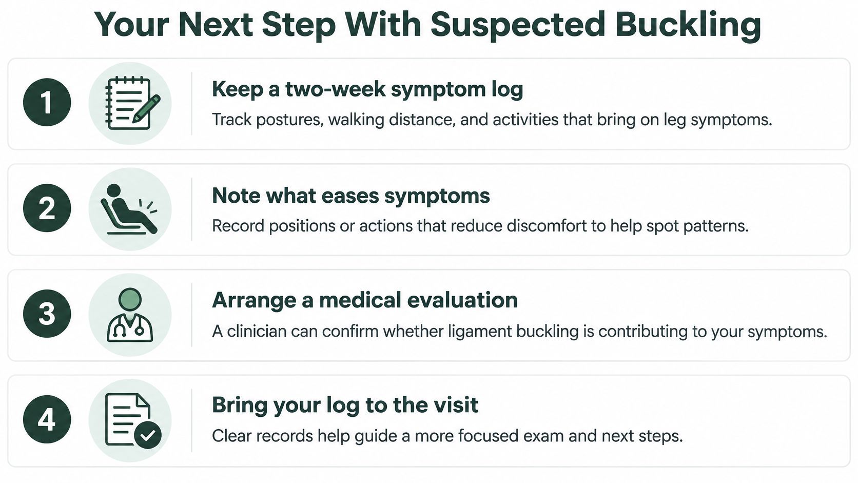
- Keep a two-week symptom log. Record which postures, walking distances, and activities bring on leg symptoms, because a fluctuating pattern is the clearest clue that narrowing may be dynamic.
- Note what eases symptoms. Write down whether sitting, leaning forward, or resting helps, and how long relief takes, since that response helps separate extension-sensitive narrowing from constant compression.
- Bring the log and your imaging report to the visit. Ask whether your images were obtained lying down or in extension, because the position shapes what the scan could and couldn't show.
- Ask whether a dynamic or upright study would clarify the picture. This question matters most when your symptoms follow standing or extension but your supine scan looks mild or doesn't match.
- Ask how the imaging level and side match your examination. Imaging alone doesn't decide treatment, so strength, sensation, and reflex findings need to point to the same level.
- Check yourself against the urgent patterns. Seek emergency care at once for progressive weakness or bladder, bowel, or saddle-area changes, rather than waiting to discuss them at a routine visit.
Choosing who to see first
For a first review, your primary care clinician can examine you, screen for warning signs, and coordinate imaging. A spine specialist becomes especially helpful when the imaging and examination don't agree, when weakness is progressing, or when you're weighing an injection or surgery. For mechanical symptoms without red flags, you can discuss a conservative plan built around movement retraining and, where suitable, osteopathic care, with clear checkpoints for whether it's helping.
The goal isn't a single answer from one scan. It's a clear mechanical diagnosis, what's folding, in which position, at which level, and with what effect on your nerves, followed by a plan matched to that diagnosis. A thick ligament on a report is the start of that conversation, not the end of it.
If your symptoms follow a pattern of standing, walking, or arching back, and your scan doesn't seem to explain them, Restore Osteopathic Medicine in Aurora offers physician-led osteopathic evaluation that compares your imaging with how your spine moves and functions. You'll get a clear explanation of what your findings do and don't mean, plus a conservative plan matched to the mechanical problem, with monitoring for any change that needs specialist referral. Visit Restore Osteopathic Medicine to schedule a consultation and bring your symptom log and imaging report with you.